サンプル調製例(Saturation dyes)
Saturation dyesを使用した2Dパターンおよびサンプル調製例(タンパク質抽出法、標識の方法、二次元電気泳動のプロトコール)を以下に示します。
Sample Types
Mouse brain
| Extraction and Labelling Protocols |
Extraction
Tissue washed with saline (0.9 %), mechanically homogenised in cell lysis buffer (7 M urea, 2 M thiourea, 4 % CHAPS, 40 mM Tris, pH 8.0, 1 ml per 0.1 g tissue) and centrifuged (13,000 rpm, 10 min, 4 ℃). Pellet discarded and supernatant used for labelling.
Labelling
5 μg protein labelled with 4 nmol TCEP and 8 nmol dye. |
| 1st and 2nd Dimension Conditions |
1st Dimension
pH 3-10 NL, 24 cm Immobiline™ DryStrips.
Ettan™ IPGphor™ IEF unit, anodic cup
loading.
50 μA per strip
1. 300 V, 3 h, step
2. 600 V, 3 h, gradient
3. 1,000 V, 3 h, gradient
4. 8,000 V, 3 h, gradient
5. 8,000 V, 4 h, step
2nd Dimension
12.5 % Ettan™ DALT gel,
2 W per gel overnight, 15 ℃. |
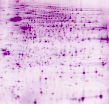
Cy3/Cy5 overlay for 5 μg protein labelled with CyDye™ DIGE Fluor Cy3 saturation dye (red) and 5 μg protein labelled with CyDye™ DIGE Fluor Cy5 saturation dye (blue).
HEP G2 cultured cell line
| Extraction and Labelling Protocols |
Extraction
Serum-free medium was poured off and cells washed twice with PBS in the flask. Without trypsinisation, cell lysis buffer (2 M thiourea, 7 M urea, 4 % CHAPS, 40 mM Tris, pH 8.0) was
added to the flask. Cell lysate was pipetted out and sonicated on wet ice, with low-intensity 30 s pulses until the lysate turned clear. The sample was centrifuged (13,000 rpm, 10 min, 4 ℃), the pellet discarded and the supernatant used for labelling.
Labelling
5 μg protein labelled with
2 nmol TCEP and 4 nmol dye. |
| 1st and 2nd Dimension Conditions |
1st Dimension
pH 3-10 NL, 24 cm Immobiline™ DryStrips.
Ettan™ IPGphor™ IEF unit, anodic cup loading.
50 μA per strip
1. 300 V, 3 h, step
2. 600 V, 3 h, gradient
3. 1,000 V, 3 h, gradient
4. 8,000 V, 3 h, gradient
5. 8,000 V, 4 h, step
2nd Dimension
12.5 % Ettan™ DALT gel,
2 W per gel overnight, 15 ℃. |
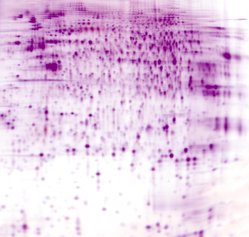
Cy3/Cy5 overlay for 5 μg protein labelled with CyDye™ DIGE Fluor Cy3 saturation dye (red) and 5 μg protein labelled with CyDye™ DIGE Fluor Cy5 saturation dye (blue).
Rat liver
| Extraction and Labelling Protocols |
Extraction
Tissue washed 4× with saline (0.9 %) and mechanically homogenised in cell lysis buffer (8 M urea, 4 % CHAPS, 30 mM Tris, pH 8.0, 1 ml per 0.1 g tissue). The supernatant was extracted and sonicated on wet ice, with low-intensity 30 s pulses until the lysate turned clear. The sample was centrifuged (13,000 rpm, 10 min, 4 ℃), the pellet discarded and the supernatant used for labelling.
Labelling
5 μg protein labelled with
2 nmol TCEP and 4 nmol dye |
| 1st and 2nd Dimension Conditions |
1st Dimension
pH 3-10 NL, 24 cm Immobiline™ DryStrips.
Ettan™ IPGphor™ IEF unit, anodic cup loading.
50 μA per strip
1. 300 V, 3 h, step
2. 600 V, 3 h, gradient
3. 1,000 V, 3 h, gradient
4. 8,000 V, 3 h, gradient
5. 8,000 V, 4 h, step
2nd Dimension
12.5 % Ettan™ DALT gel,
2 W per gel overnight, 15 ℃. |
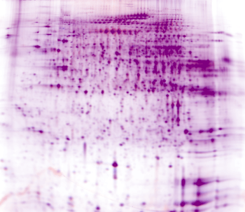
Cy3/Cy5 overlay for 5 μg protein labelled with CyDye™ DIGE Fluor Cy3 saturation dye (red) and 5 μg protein labelled with CyDye™ DIGE Fluor Cy5 saturation dye (blue).
Rat lung
| Extraction and Labelling Protocols |
Extraction
Tissue washed 4× with saline (0.9 %) and mechanically homogenised in cell lysis buffer (8 M urea, 4 % CHAPS, 30 mM Tris, pH 8.0, 1 ml per 0.1 g tissue). The supernatant was extracted and sonicated on wet ice, with low-intensity 30 s pulses until the lysate turned clear. The sample was centrifuged (13,000 rpm, 10 min, 4 ℃), the pellet discarded and the supernatant used for labelling.
Labelling
5 μg protein labelled with
2 nmol TCEP and 4 nmol dye. |
| 1st and 2nd Dimension Conditions |
1st Dimension
pH 3-10 NL, 24 cm Immobiline™ DryStrips.
Ettan™ IPGphor™ IEF unit, anodic cup loading.
50 μA per strip
1. 300 V, 3 h, step
2. 600 V, 3 h, gradient
3. 1,000 V, 3 h, gradient
4. 8,000 V, 3 h, gradient
5. 8,000 V, 4 h, step
2nd Dimension
12.5 % Ettan™ DALT gel,
2 W per gel overnight, 15 ℃. |
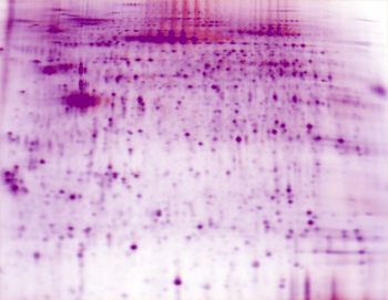
Cy3/Cy5 overlay for 5 μg protein labelled with CyDye™ DIGE Fluor Cy3 saturation dye (red) and 5 μg protein labelled with CyDye™ DIGE Fluor Cy5 saturation dye (blue).
お問合せフォーム
※日本ポールの他事業部取扱い製品(例: 食品・飲料、半導体、化学/石油/ガス )はこちらより各事業部へお問い合わせください。
お問い合わせありがとうございます。
後ほど担当者よりご連絡させていただきます。
|
